


")


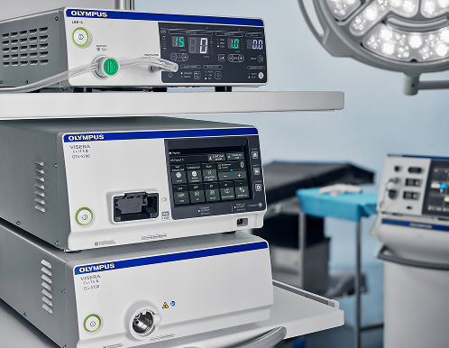
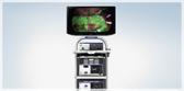
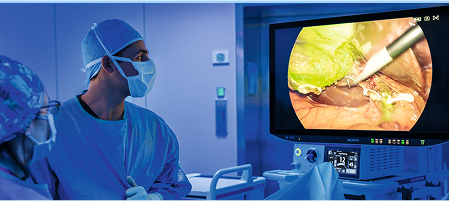
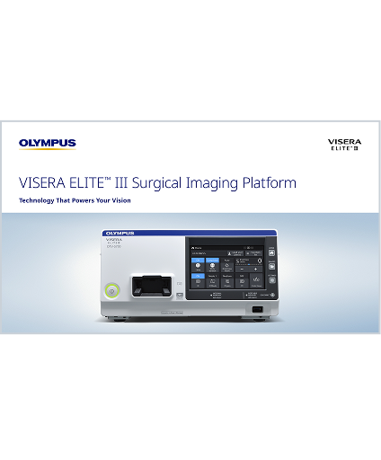
The VISERA ELITE™ III Multispecialty
Surgical Imaging Platform
Technology That Powers Your Vision
The VISERA ELITE™ III platform delivers imaging centered around:
- Light — Bright Image
- Color — More Colors1
- Clarity — Never Lose Focus2
VISERA ELITE™ III surgical imaging platform was designed by Sony and Olympus (SOMED) to balance light, color, and clarity in a future ready, multispecialty, 4K/3D advanced imaging platform. Backward compatibility* and software upgrades may help hospitals reduce upfront costs while accessing the latest technology for patient care.
*See IFU for full list of compatible scopes


NBI™ Technology is not intended to replace histopathological sampling as a means of diagnosis.
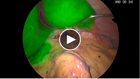
Fluorescence Imaging with the
VISERA ELITE™ III Platform
The VISERA ELITE III platform surgical supports infrared (IR) observation with indocyanine green (ICG) to assist intraoperative visualization of key anatomical and physiological features.
Infrared (IR)
- Perfusion & Vascular Flow: Visualizes blood flow and ischemic areas.
- Anastomotic Evaluation: May help reduce leakage during esophageal and colorectal procedures.3
- Organ Visualization: Enables imaging of bile ducts, kidneys, lymphatics, and more.
Customize your IR Image
Adjust the visibility of the fluorescence by controlling IR gain to meet the best visualization regardless of scope, tissue or environment changes. The IR gain can be adjusted in three levels: low, medium and high. The higher the level, the stronger the fluorescence is expressed.
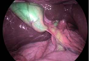
Full-color IR White-light Overlay Mode
Providing real-time 4K fluorescence imaging during white-light observation.
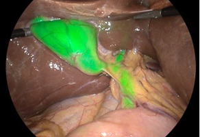
IR Magenta Mode
Regular tissue shown in magenta, while ICG appears green. These two colors are the complete opposite on the color wheel resulting in the highest contrast image for greater clarity.
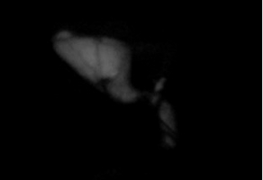
IR Black & White Only Mode
High contrast image of fluorescence displayed in grayscale.
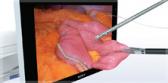
Advancing Surgical Visualization
In a 2025 survey, 8 out of 10 surgeons surveyed agreed that YE may assist in identifying vessels, nerves, and critical structures within adipose tissue.4
Yellow Enhancement (YE)
Yellow Enhancement (YE) is a processor-based imaging mode in the VISERA ELITE™ III platform that enhances the visibility of yellow-containing structures, such as fatty tissue, by converting pale orange tones into vivid yellow. This contrast supports identification of critical anatomy without the need for dyes, drugs, or specialized scopes.
This mode works by converting the orange color of adipose tissue containing beta-carotene into high contrast yellow.

Our Latest-generation Multispecialty
Surgical Imaging Platform
Optimized Investment Tailored to Your Needs
Software-upgradable platform designed to adapt to evolving OR requirements.
Advancing Surgical Visualization
Designed to support surgical visualization across specialties by combining technologies like 4K, 3D, Narrow Band Imaging™ (NBI™) Technology, and Yellow Enhancement (YE) mode.
All-in-One Multispecialty Platform
Support for over a dozen different video and rigid endoscopes across general surgery, bariatric, colorectal, thoracic, urology, gynecology, airway, and more.
See IFU for the full list of compatible devices.
Never Lose Focus
Experience sharp images through True 4K CMOS sensor and constantly keep the focus with Continuous Auto Focus (CAF) function and Extended Depth of Field (EDOF) technology.2
One System for All
Designed to meet the needs of multiple specialties including general surgery, urology, gynecology, ENT and more, the VISERA ELITE™ III multispecialty surgical imaging platform offers 3D, IR, and 4K imaging, Narrow Band Imaging™ (NBI™) Technology* and Yellow Enhancement (YE) mode, all in one system. Compatibility with flexible ENT and urology flexible videoscopes, camera heads, and ENDOEYE™ videoscopes allows you to adjust your investment for efficiency.**
* NBI™ Technology is not intended to replace histopathological sampling as a means of diagnosis.
** Please refer to the IFU for compatible devices.
-
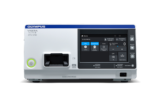
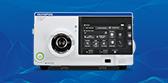
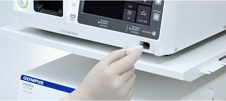
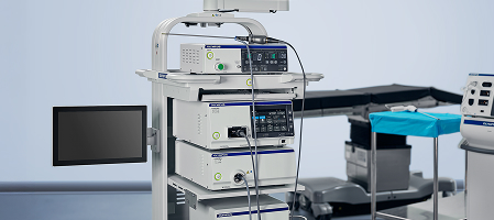
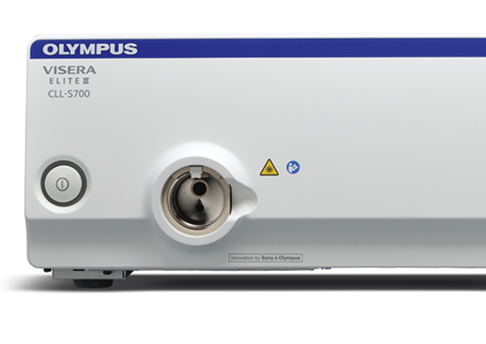
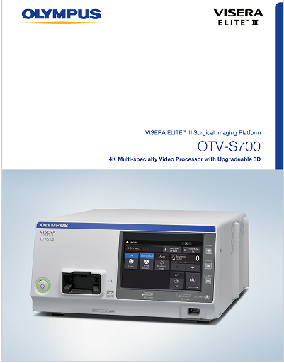
OTV-S700 Processor
VISERA ELITE III Multispecialty
Surgical Imaging PlatformAs an all-in-one system that supports your needs, the VISERA ELITE III platform can be upgraded via software licensing when needed — helping to streamline workflows and minimize OR downtime for the upgrade process.
VISERA ELITE III is a compact, multispecialty imaging platform designed for laparoscopic diagnosis, treatment, and video observation. TYPE BF endoscopes connected to this imaging platform must never be applied directly to the heart. Leakage current from the TYPE BF applied part may be dangerous and cause ventricular fibrillation or otherwise seriously affect the cardiac function of the patient.
-
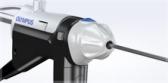
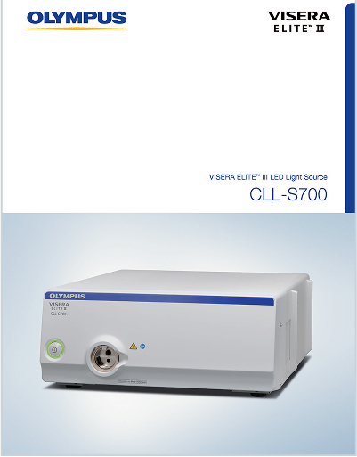
VISERA ELITE III LED Light Source
VISERA ELITE™ III LED light source is designed to provide natural color reproduction using long-life LED bulbs, supporting operational efficiency over time.
-
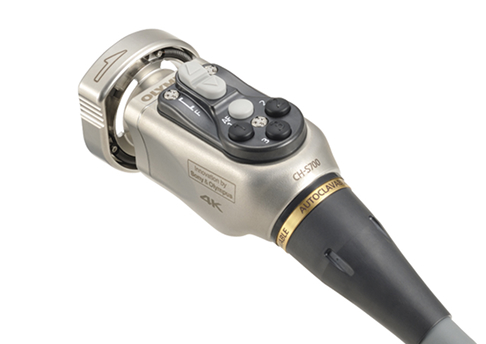
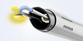
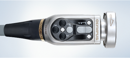
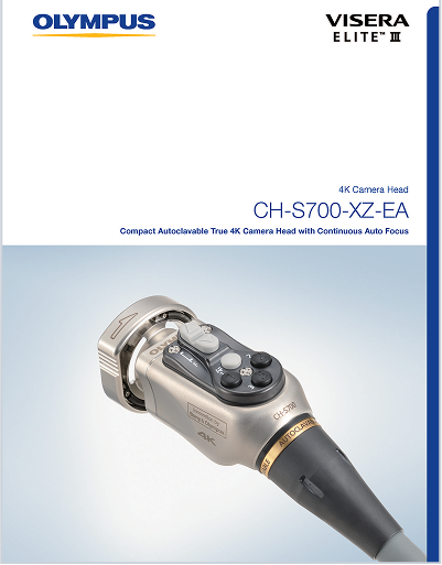
4K Camera Head
Discover the details in true 4K image quality with the small and lightweight camera head featuring NBI™ Technology and Yellow Enhancement (YE) observation modes. The new Continuous Auto Focus (CAF) function helps to minimize distractions and Extended Depth of Field (EDOF™) technology facilitates endoscopic observations through continuous broad focus and enhanced depth of field allowing more of the image to be in focus at once.3
-
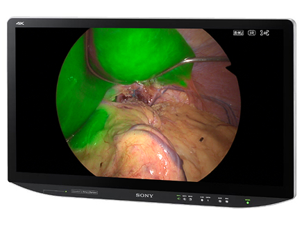
3D 4K Medical LCD Monitor
(Available in 32" and 55")
Experience high-quality 4K UHD video images in 3D and 2D with a High Dynamic Range (HDR) setting.
-
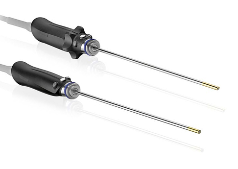
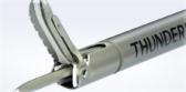
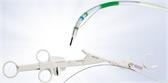
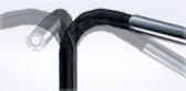
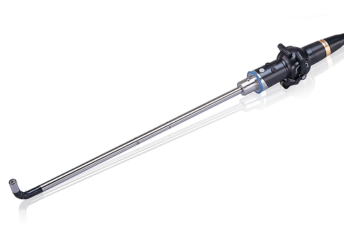
ENDOEYE™ Rigid Videoscope
The ENDOEYE Rigid video laparoscope combines vivid visualization with one-handed image rotation, allowing the surgeon to adjust the view direction while maintaining a stable horizon. Its advanced chip-on-tip technology is designed to deliver bright, clear, and focus-free images. Select models also feature integrated fog-resistant elements to help reduce scope fogging during procedures.
ENDOEYE has been designed to be used within the thoracic and abdominal cavities including female reproductive organs. This instrument must not be used for observation or treatment of the heart and must not contact the heart or any area near the heart or contact any device or therapeutic accessory that contacts the heart or any area near the heart. Do not use this instrument for any purpose other than its intended use.
-
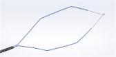
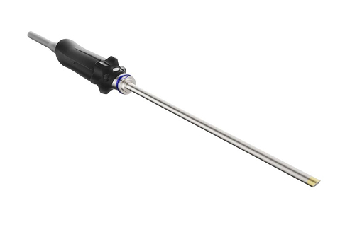
ENDOEYE™ FLEX Videoscope
Shift the field of view with ENDOEYE FLEX videoscope 5 mm and 10 mm by controlling the tip angulation up to 100° to the desired location. With access to narrow cavities, obtain a clear viewing angle of structures in areas such as the rectum, VATS procedures, pelvic cavity, retroperitoneal approach to urology, etc.
ENDOEYE has been designed to be used within the thoracic and abdominal cavities including female reproductive organs. This instrument must not be used for observation or treatment of the heart and must not contact the heart or any area near the heart or contact any device or therapeutic accessory that contacts the heart or any area near the heart. Do not use this instrument for any purpose other than its intended use.
-
ENDOEYE™ Rigid 3D Videoscope
The autoclavable, rigid ENDOEYE 3D videoscope provides natural 3D depth perception with a wide field of view.
Change the direction of view while maintaining a stable horizon. The ENDOEYE Rigid 30° videoscope supports your continuous critical view and provides you a reliable orientation even at challenging viewing angles with its continuous mechanical rotation function.
ENDOEYE has been designed to be used within the thoracic and abdominal cavities including female reproductive organs. This instrument must not be used for observation or treatment of the heart and must not contact the heart or any area near the heart or contact any device or therapeutic accessory that contacts the heart or any area near the heart. Do not use this instrument for any purpose other than its intended use.
-
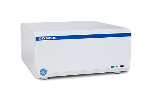
nCare™ 4K Medical Recorder
Secure, intuitive, and designed to support healthcare teams, the nCare 4K recorder is a network connected medical recorder that can capture 4K and/or HD images and videos from up to two devices simultaneously.
-
.jpg)
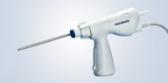
Intraabdominal Insufflation Unit (UHI-4)
The Olympus® UHI-4 i Intraabdominal insufflation unit is designed to insufflate the abdominal cavity during diagnostic and operative laparoscopic procedures. The system also includes smoke evacuation functionality and a small cavity mode. Automatic smoke evacuation is available when used in combination with Olympus® ESG or USG electrosurgical generators.
Discover the Power of the
VISERA ELITE™ III Platform
Experience the next generation of multispecialty surgical imaging designed to elevate precision and efficiency in the OR. See it in action with a live demonstration and get expert guidance from an Olympus® consultant

Never Lose Focus
Continuous Auto Focus (CAF)
The CAF function maintains constant focus, eliminating the need for manual adjustments and it is designed to support surgeon comfort and reduce fatigue throughout the surgery.5
Extended Depth of Field (EDOF™) Technology
EDOF technology allows more of the image to be in focus at once.1
True 4K Image Quality
Sony’s exclusive 4K sensor delivers exceptional detail for the intricate surgical procedures.
Narrow Band Imaging™ (NBI™) Technology
NBI™ Technology enhances visibility of vascular structures on the mucosal surface.
NBI™ Technology is not intended to replace histopathological sampling as a means of diagnosis.
3D imaging can go beyond the limits of what traditional endoscopes can see.
Olympus® 3D systems have been shown to offer enhanced depth perception and a precise spatial view of anatomy that simply cannot be achieved with traditional 2D systems.6
3D imaging provides:
- Enhanced Depth Perception6
- Improved Speed7
- Improved Accuracy & Precision8
Medical Expert Training
Hands-on courses, e-learnings, workshops, peer-to-peer trainings, accredited continuing trainings, and custom on-demand learning for physicians who want to develop their skills and knowledge.
Browse through our lists and find the education that fits your needs.

Are you interested in experiencing the VISERA ELITE™ III multispecialty surgical imaging platform?
Request more information or a live demonstration and an Olympus® consultant will contact you.
VISERA ELITE III is a compact, multispecialty imaging platform designed for laparoscopic diagnosis, treatment, and video observation. TYPE BF endoscopes connected to this imaging platform must never be applied directly to the heart. Leakage current from the TYPE BF applied part may be dangerous and cause ventricular fibrillation or otherwise seriously affect the cardiac function of the patient.
- Data on file with Olympus as of 04/2025.
- Continuous Autofocus (CAF) is designed to automatically maintain image focus and works in conjunction with Extended Depth of Field (EDOF™) technology to maximize the in-focus area.
- Faber RA, Meijer RPJ, Droogh DHM, Jongbloed JJ, Bijlstra OD, Boersma F, Braak JPBM, Meershoek-Klein Kranenbarg E, Putter H, Holman FA, Mieog JSD, Neijenhuis PA, van Staveren E, Bloemen JG, Burger JWA, Aukema TS, Brouwers MAM, Marinelli AWKS, Westerterp M, Doornebosch PG, van der Weijde A, Bosscha K, Handgraaf HJM, Consten ECJ, Sikkenk DJ, Burggraaf J, Keereweer S, van der Vorst JR, Hutteman M, Peeters KCMJ, Vahrmeijer AL, Hilling DE. Indocyanine green near-infrared fluorescence bowel perfusion assessment to prevent anastomotic leakage in minimally invasive colorectal surgery (AVOID): a multicentre, randomised, controlled, phase 3 trial. Lancet Gastroenterol Hepatol. 2024 Oct;9(10):924-934. doi: 10.1016/S2468-1253(24)00198-5. Epub 2024 Aug 13. PMID: 39151436.
- Data on file with Olympus (Yellow Enhancement Claims Evaluation, June 13, 2025). N=98 surgeons surveyed.
- Compared to CH-S400-XZ-EA/EB data on file with Olympus N=9 (DC00677791)
- Gabrielli M, et al “Assessment of 3-Dimensional vs 2-Dimensional Imaging and Technical Performance Using a Multiport Intraoperative Data Capture and Analytic System for Patients Undergoing Laparoscopic Roux-en-Y Gastric Bypass Surgery” JAMA Netw Open 2020; DOI: 10.1001/jamanetworkopen.2019.20084.
- Data on File with Olympus as of April 2020. Claims based on one study.